


0 Introduction
Brassinosteroids, a type of traditional phytohormone, are widely exist in a various plants. In 1979, brassinolide was identified as the most representative brassinosteroid isolated from rape pollen. It plays an crucial role in plant differentiation, disease resistance, root growth, stress tolerance, and the aging process of plants, etc[1]. Brassinolide is most commonly found in the pollen, seeds, roots, stems, fruits, and leaves of many plants[2]. The concentration of brassinolide in plants is typically very low, ranging from 0.01 to 100 ng/g FW (Fresh weight)[3]. Therefore, a sensitive and accurate method for detecting brassinolide is necessary.
The conventional detection techniques for brassinosteroids include bioassay[4], chromatography[5] and immunoassay (such as radioimmunoassay and enzyme-linked immunosorbent assay)[6], as well as liquid chromatograph-mass spectrometer[7]. However, these detection methods are all conducted in vitro. They require expensive equipment, complex preprocessing procedures, highly skilled operators, or extended operating times. With the rapid development of precision agriculture, there is a growing interest among researchers in acquiring real-time, in situ and in vivo plant physiological information. Therefore, new methods for detecting brassinolide need be developed.
Electrochemical sensors have been widely utilized for the detection of various substances, including proteins[8], nucleic acids[9], small molecules in plants[10], and ions[11], owing to their simple operation, rapid response, cost-effective instrumentation, and superior detection sensitivity. Electrochemical immunosensors can utilize the high selectivity of antibodies and the high sensitivity of electrochemical sensors, and hold potential for in situ detection of biomolecules. However, as of now, there are no reports on the use of electrochemical immunosensors for the detection of brassinolide.
Two-dimensional (2D) materials, such as Mxene, stand out due to their unique metallic conductivity, high specific surface area, and numerous functional groups. These advantageous characteristics have led to the widespread use of Mxene in the fields of energy storage[12], biotherapeutic[13, 14], electrocatalytic water splitting[15], sensors[16, 17], and more. However, the strong van der Waals force between adjacent sheets make is easy to produce densely packed lamellas, limiting its practical application[13, 14, 18]. To address this, researchers have explored the use of different nanomaterials as additives for interlayer of Mxene, such as MWNTs and GO. Additionally, dopamine (DA), a small and low-cost organic molecule, can spontaneously polymerize and synthesize polydopamine (PDA) films on various surfaces under weakly alkaline conditions[19]. Moreover, the high content of Catechol in PDA provides robust adhesive properties, making it particularly advantageous for coating nanomaterials, generating nano coatings and altering their surface properties[10]. PDAs can also serve as binders and crosslinkers, not only to immobilize inorganic nanoparticles, but also to further functionalize polymers for enhanced adsorption capacity. For instance, Deng et al.[20] prepared PDA@Mxene as printing ink, leveraging PDA to enhance the waterproof and antioxidant properties of Mxene. Similarly, Lee et al.[21] employed DA to create an adhesive layer by in situ polymerization on MXene, resulting in a composite material with a highly organized and tightly layered structure that not only improves the effective shielding against oxygen and moisture, but also significantly improves the stability of Mxene. However, the potential application properties of Mxene@PDA in biosensor construction requires further exploration.
In this study, AuNPs were innovatively electrodeposited onto the Screen-printed electrode (SPE) due to their excellent conductivity. CuCl2 NWs, as a one-dimensional (1D) nanomaterial, not only exhibit exceptional conductivity and chemical stability, but also can be easily obtained by simple synthetic methods. Additionally, the redox peak of Cu2+ can also serve as an electrical signal probe for the electrochemical immunosensor, eliminating the need for an additional redox probe in electrolytic solutions, as required in traditional immunosensors. Consequently, CuCl2 NWs were employed to modify the electrode. Furthermore, Mxene@PDA nanocomposite was coated on the electrode to enhance its conductivity and biocompatibility. Upon modification with brassinolide monoclonal antibodies, the resulting immunosensor demonstrated high sensitivity and selectivity in detecting brassinolide. It was also successfully utilized for in situ detection of brassinolide in salt-treated wheat samples.
1 Materials and methods
1.1 Chemicals and materials
Monoclonal antibodies against brassinolide were purchased from China Agricultural University. Brassinolide standards (C28H48O6) and Thiourea were sourced from Shanghai Macklin Biochemical Technology Co., Ltd. (Shanghai, China). Anhydrous cupric chloride (CuCl2), Bovine serum albumin (BSA) and gold chloride trihydrate (HAuCl4·3H2O) were provided by Sigma-Aldrich (St. Louis, USA). Ti3C2 MXene nanosheets were obtained from 11 Technology Co., Ltd. (Jilin, China). Tris (hydroxymethyl) aminomethane (C4H11NO3) was purchased from Beijing Biodee Biotechnology Co., Ltd. Brassinolide was diluted with phosphate buffer solution (PBS, 0.01 M, pH 7.4).
1.2 Instruments
The surface morphology of the modified sensor was analyzed using a FESEM (Field emission scanningelectron microscope) system (ZEISS, SEM 500) equipped with an EDS (Energy Dispersive Spectrometer) detector. A 5 mm diameter glassy carbon sheet served as the substrate for emission scanningelectron microscope (SEM) and EDS mapping. X-ray photoelectron spectroscopy (XPS) experiments were carried out using an AXIS HIS 165 spectrometer of Kratos Analytical. The SPEs, consisting of a carbon-based working electrode (d=2.5 mm), a Ag/AgCl reference electrode, and a carbon-based counter electrode, were supplied by Ningbo Mxense Biotechnology Co., Ltd. All electrochemical experiments were performed using an electrochemical CHI760E workstation (Shanghai Chenhua Instrument Co., Ltd., China).
1.3 Synthesis of CuCl2 NWs and Mxene@PDA
Copper(II) chloride nanowires (CuCl2 NWs) were synthesized following a previous reported method[22]. In brief, 0.05 g CuCl2, and 0.05 g thiourea were mixed with 40 mL of ethanol. After sonicating the mixture for 5 min, it was centrifugated at 3 000 r/m for 5 min. Subsequently, the resulting material was rinsed and dried to obtain the CuCl2 NWs powder.
The Mxene@PDA nanocomposite was prepared following a previous published method[23]. Initially, 0.01 g of Tris (C4H12NO3) was added to a dispersion of Mxene (0.5 mg/mL). After sonicating for 5 min, the mixture was adjusted to a weakly basic pH. Subsequently, 0.005 g of DA was added and stirred at room temperature in the dark for 24 h, resulting in the formation of a Mxene@PDA suspension. The product was washed 3 to 5 times with ethanol and deionized water. After drying, the precipitate was collected, yielding the Mxene@PDA composite material.
1.4 Construction of electrochemical immunosensor
Fig. 1 depicts the preparation procedure of the immunosensor. Initially, AuNPs were electrodeposited onto the cleaned SPE surface using the IT method (-1.3 V, 600 s) in a PBS solution containing 0.6 g/mL of HAuCl4, resulting in the modified electrode noted as AuNPs/SPE. Subsequently, 4 µl of CuCl2 NWs (diluted to 0.2 mg/mL with deionized water) was dropped onto the SPE and dried in a 37 ℃ oven, leading to the modified electrode defined as CuCl2 NWs/AuNPs/SPE. Following this, 4 µL of the Mxene@PDA suspension was dropped on the electrode, yielding the electrode denoted as Mxene@PDA/CuCl2 NWs/AuNPs/SPE. Then, the brassinolide monoclonal antibody (0.08 mg/mL) was incubated with the electrode for 1 h at room temperature, resulting in the electrode defined as antibody-brassinolide/Mxene@PDA/CuCl2 NWs/AuNPs/SPE. Finally, 2 µL of BSA (1 wt%) was added to the electrode to block the nonspecific binding site. After drying at room temperature for 30 min (24 °C), the obtained electrode was defined as BSA/antibody-brassinolide/Mxene@PDA/CuCl2 NWs/AuNPs/SPE.
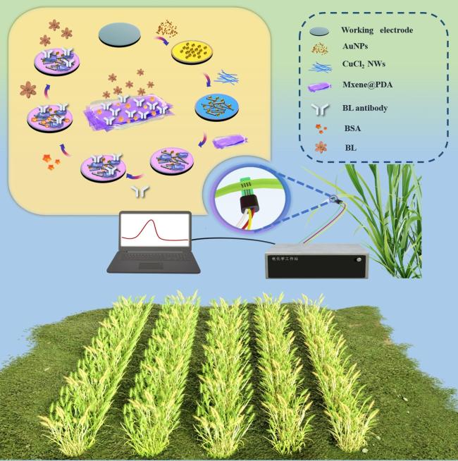
Fig. 1 Schematic illustration of the fabrication process of the electrochemical immunosensor for brassinolide |
1.5 Measurement procedures
The modification procedure of the proposed immunosensor was investigated using electrochemical impedance spectroscopy (EIS), which was conducted with frequency parameters ranging from 1×10⁻² to 1×10⁵ Hz and voltage amplitudes of 5 mV. The performance of the immunosensor was evaluated by differential pulsed voltammetry (DPV), with a potential range of -0.5 to 0.5 V (Pulse duration: 0.2 s; Pulse amplitude: 50 mV; Pulse width: 50 ms). A PBS buffer (0.01 M, pH 7.4) served as the electrolyte for brassinolide detection. Brassinolide standard solutions of varying concentrations were individually applied to the SPE and then incubated for 1 hour. Following each incubation, the sensor was rinsed with PBS buffer to remove any unfixed materials.
1.6 Preparation of planting material
The wheat variety tested in the experiment, Cangmai 6005, was obtained from the National Precision Agriculture Experimental Station in Beijing, China. The seeds were sterilized and soaked before being immersed in distilled water and placed in a light incubator for 16 h to facilitate seed germination (light/dark period: 10 h/14 h, temperature: 25 ± 2 ℃, humidity: 60%). The germinated seedlings were transplanted into Hoagland's solution. Wheat seedlings served as the raw material for the recovery determination. The wheat samples were ground in liquid nitrogen and dissolved in PBS buffer. Subsequently, the samples were refrigerated overnight at 4 °C, followed by centrifugation (12 000 r/m, 8 min). The resulting supernatant was collected and used for the recovery test.
Twenty wheat plants with similar growth were randomly divided into two groups, with 10 plants in each group. The control group was treated with Hoagland's solution, while the other group was treated with Hoagland's solution containing 100 mM NaCl. After 4 days of treatment, in situ tests were carried out. Small holes were punctured in the surface of the wheat leaves using a puncture tool. The prepared sensor was fixed to the tested area with tape. Subsequently, 20 µL of PBS buffer was added to the small holes, allowing the sap of the wheat leaves to flow out and comes into contact with the working electrode, followed by the DPV test.
2 Results and discussion
2.1 Characterization of the immunosensor
The electrode's morphology was investigated using SEM. The bare SPE exhibited a clean and smooth surface, as depicted in Fig. 2(a). Subsequently, AuNPs were electrodeposited on the electrode, uniformly covering the surface with particle measuring 70–100 nm (Fig. 2(b)). This deposition enhances the electrode's conductivity and effective active area due to the excellent conductivity and large specific surface area of AuNPs. Following this, CuCl2 NWs were applied to the surface of the AuNPs, revealing a linear structure of CuCl2 laid on the granular structure of the AuNPs (Fig. 2(c)). Upon modifying the electrode with the Mxene@PDA composite material, a membrane structure was observed (Fig. 2(d)), which serves to enhance the electrode's conductivity, biocompatibility and antibody adsorption ability. Upon the addition of brassinolide antibodies to the electrode's surface, numerous macromolecules were observed covering the electrode, originating from antibody molecules or antibody aggregation, indicating the successful modification of brassinolide antibodies on the electrode (Fig. 2(e)). Lastly, when BSA was introduced to the electrode (Fig. 2(f)), the protein molecules exhibited increased aggregation and formed a film structure, signifying the immobilization BSA molecules on the electrode.
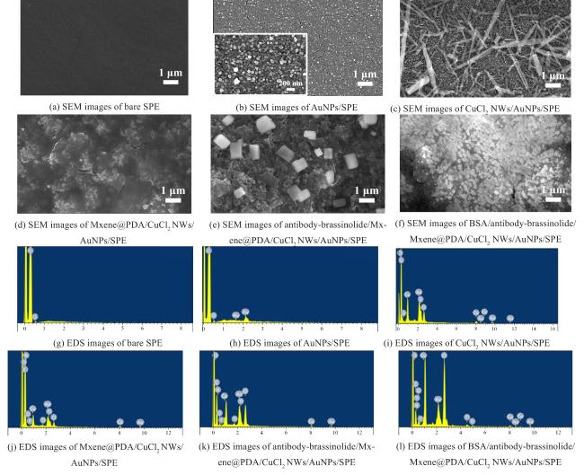
Fig. 2 SEM and EDS images of immunosensor |
Elemental analysis of the modified electrode was performed using the EDS technique (Fig. 2(g)~ Fig. 2(l)). The working electrode of the SPE was carbon-based, and the EDS results of the bare SPE revealed the presence of the element C (Fig. 2(g)). Following the plating AuNPs on the electrode, the EDS results also showed the presence of the Au element (Fig. 2(h)). In the case of CuCl2 NWs/AuNPs/SPE, the EDS result also included Cu and Cl elements, primarily due to the modification of CuCl2 NWs material (Fig. 2(i)). For Mxene@PDA/CuCl2 NWs/AuNPs/SPE, the EDS analysis also presented Ti, F and N elements. The presence of Ti was attributed to the modification of Mxene, while the presence of F was linked to the fact that Mxene (Ti3C2) was etched in concentrated hydrofluorine acids (HF) before purchase. The presence of N primarily due to the fact that N is the main element of PDA (Fig. 2(j)). Since the main element in proteins is N, no additional elements appeared after the immobilization of protein molecules (antibody and BSA) on the electrode (Fig. 2(k) and Fig. 2(l)). The results of SEM and EDS mapping confirm the successful construction of the immunosensors.
Mxene and Mxene@PDA were analyzed by XPS to investigate the binding between PDA and Mxene. A peak of N 1 s (401.1 eV) was detected in Mxene@PDA (Fig. 3(a), Curve b), confirming the effective introduction of PDA into MXene. The spectra of C 1 s of Mxene@PDA, as shown in Fig. 3(b), exhibited six major peaks at 281.6 (C-Ti), 284.2 (C-NH2), 284.9 (C-C), 286.1 (C-O/C-N), 286.8 (C-O-Ti), and 289.1 eV (C=O), respectively [24]. The presence of a peak associated with the C-O-Ti bond at 286.8 eV in the C 1 s spectrum of Mxene@PDA indicated that Mxene and PDA were linked to each other by catechol-titanium coordination bonds [21]. Furthermore, four characteristic peaks of N 1 s were observed in Mxene@PDA (Fig. 3(c)). The peak for R-NH2 (400.88 eV) is indicative of DA, while the peak for R-NH-R (399.79 eV) is related to PDA or oxidized intermediates [25]. It has been reported that the tautomers of 5,6-indolequinone and 5,6-dihydroxyindole were responsible for the =N-R peak (398.3 eV) [23]. Additionally, a new peak of the N-Ti bond was observed at 395.8 eV[26]. The O 1 s spectrum of the Mxene@PDA revealed a C-O-Ti peak (Fig. 3(d)), indicating that Mxene and PDA were coupled through a coordination bond[27]. These findings confirm that PDAs can serve as binders or crosslinkers to closely arrange adjacent nanosheets of Mxene.
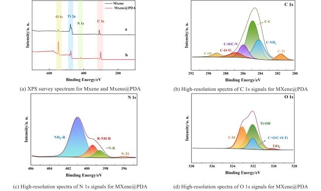
Fig. 3 High-resolution XPS spectra of Mxene@PDA showing the chemical composition and bonding structures |
2.2 Feasibility of immunosensor for detecting brassinolide
The feasibility of the immunosensor for detecting brassinolide was assessed using EIS and DPV methods. As shown in Fig. 4(a), the diameter of the semicircle represented the electron transfer resistance (Rct). The Rct of the bare SPE electrode was relatively large (Rct=3.75 kΩ) (Fig. 4(a), Curve a). Upon deposition of AuNPs onto the working electrode of SPE (Fig. 4(a),Curve b), the Rct decreased dramatically to 1.12 kΩ due to the good conductivity of AuNPs. Subsequently, the Rct value decreased further with the addition of CuCl2 NWs on the surface of AuNPs/SPE(Fig. 4(a), Curve c), attributed to the good conductivity and large surface area of CuCl2 NWs. When Mxene@PDA was modified on the CuCl2 NWs/AuNPs/SPE electrode (Fig. 4(a),Curve d), Rct decreased even further to 540 Ω, indicating the excellent conductivity of Mxene@PDA. Subsequently, the continuous addition of antibody-brassinolide (Fig. 4(a), Curve e, Rct=1.48 kΩ) and BSA (Fig. 4(a), Curve f, Rct= 6.55 kΩ) on the Mxene@PDA/CuCl2 NWs/AuNPs/SPE electrode led to a gradual increase in Rct due to the insulation of these proteins, hindering electron transfer. Finally, after the binding of brassinolide (0.08 mg/mL), Rct increased further due to the insulated brassinolide molecules(Fig.4(a), Curve g). These results indicate the successful preparation of electrochemical immunosensors capable of recognizing brassinolide.
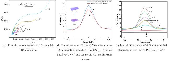
Fig. 4 Characterization experiments results of the sensor |
DPV (Differential Pulse Voltammetry) was also employed to further confirm the feasibility of the immunosensor. In Fig. 4(b), it is evident that Mxene@PDA/CuCl2 NWs/AuNPs/SPE exhibits a stronger redox peak current than Mxene/CuCl2 NWs/AuNPs/SPE, affirming the superior suitability of Mxene@PDA as a material for constructing brassinolide immunosensors. Notably, no redox peak is observed for the bare SPE electrode in the -0.2~0.4 V range (Fig. 4(c), Curve a), nor for the AuNPs modified electrode (Fig. 4(c),Curve b). Upon the modification of CuCl2 NWs on the electrode surface, an oxidation peak is observed at about 0.1 V, attributed to the redox of Cu2+ (Fig. 4(c),Curve c) [28]. Subsequently, after the deposition of Mxene@PDA dispersion, the redox peak current increases (Fig. 4(c), Curve d) owing to the excellent conductivity of MXene@PDA. As the protein molecules (antibody-brassinolide and BSA) were successively modified on the electrode, the oxidation peak of Cu2+ is progressively reduced (Fig. 4(c), Curve e and Curve f) due to their insulation characteristics. Finally, the oxidation peak of Cu2+ is further reduced (Fig. 4(c), Curve g) after the binding between antibody-brassinolide and brassinolide, indicating the insulation of brassinolide. The difference between the Cu2+ peak current before and after brassinolide binding (ΔI=IPBS–IBL) was linked to the concentration of brassinolide, enabling the quantification of the brassinolide concentration. The DPV results were consistent with EIS results, further substantiating the feasibility of the brassinolide electrochemical immunosensor.
2.3 Optimization of the immunosensor
The impact of HAuCl4 concentration on ΔI was investigated (Fig. 5(a)). As the HAuCl4 concentration was raised from 0.2 to 0.6 mg/mL, ΔI showed a significant increase. Upon using 0.6 mg/mL of HAuCl4, the value of ΔI reached its peak. However, ΔI started to decrease as the HAuCl4 concentration was further increased to 1 mg/mL. Consequently, 0.6 mg/mL HAuCl4 was chosen for subsequent experiments.
The investigation into the optimal concentration of CuCl2 NWs on the electrode revealed that, as the concentration of CuCl2 NWs increased from 1 mg/mL to 2 mg/mL, ΔI reached its maximum value. However, as the concentration of CuCl2 NWs continued to increase, ΔI subsequently decreased. Consequently, the optimal concentration of CuCl2 NWs was determined to be 2 mg/mL (Fig. 5(b)).
Next, the impact of different ratios of Mxene and DA (1:50, 2:50, 3:50, 4:50, 5:50) on ΔI was also investigated and the highest ΔI values were achieved when the ratio of Mxene and DA was 4:50 (with Mxene at 0.4 mg/mL) (Fig. 5(c)).
Finally, the impact of varying antibody concentrations on ΔI was examined. When the antibody concentration increased from 0.01 to 0.08 mg/mL, the value of ΔI showed an increasing trend. However, when the concentration of antibody was further raised from 0.08 to 0.1 mg/mL, ΔI no longer exhibited an increase (Fig. 5(d)). This observation could be attributed to the saturation of the electrode surface with the adsorbed antibodies. Therefore, the optimal antibody concentration in this study was determined to be 0.08 mg/mL.
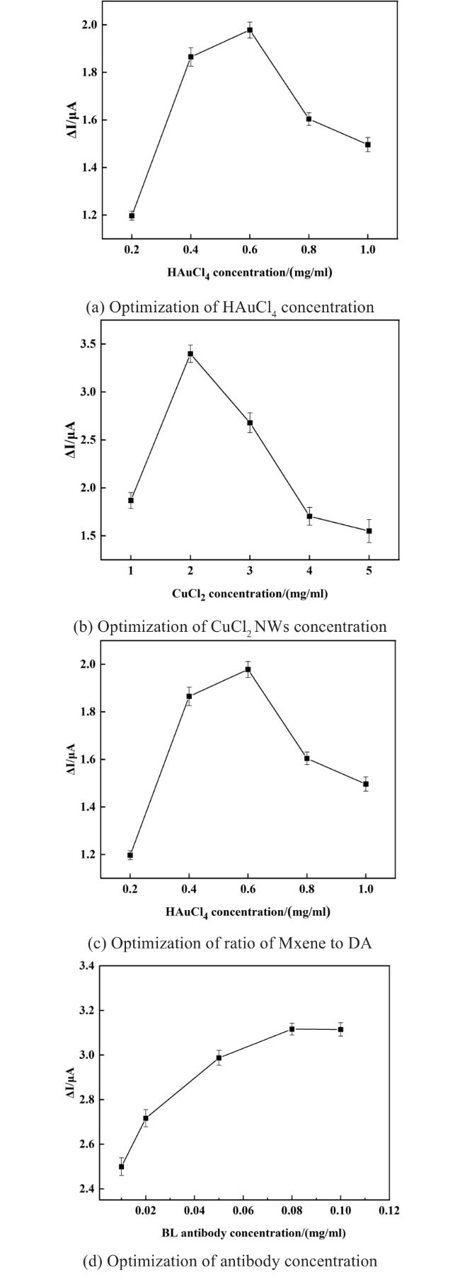
Fig. 5 The optimization of preparation conditions of brassinolide immunosensor (Error bar=SD; n=3) |
2.4 The performance of the immunosensor
The fabricated sensor was subjected to treatment with various concentrations of brassinolide ranging from 0.1pg/mL to 1 mg/mL). As illustrated in Fig. 6(a), the peak current exhibited a gradual reduction with the increment of brassinolide concentrations, which can be attributed to the insulating effect of brassinolide molecules. A highly favorable linear relationship was established between the logarithmic values of the brassinolide concentration and the corresponding ΔI values across the concentration spectrum from 0.1pg/mL to 1 mg/mL, as shown in Fig. 6(b). The limit of detection (LOD) was determined to be 0.015 pg/mL (S/N=3). The regression equation is represented by Equation (1) .
ΔI (μA)= 0.596 48l g CBL(pg/mL) + 1.540 04
The coefficient of determination (R2) is 0.998 16, indicating a very high degree of accuracy in the model. In comparison with other brassinolide detection methods (as shown in Table 1), the developted sensor boasts the broadest detection range for brassinolide, which is nearly comprehensive for the brassinolide content found in various plants, ranging from approximately 0.01 to 100ng/g of fresh weight[3]). Additionally, the sensor possesses the lowest LOD. The Mxene@PDA nanocomposite plays a pivotal role in the sensor's superior properties by offering an increased number of active sites for the antibody to bind, while also enhanceing the conductivity, stability and biocompatibility of the sensors.
Table 1 Comparison of the prepared brassinolide sensor with the previous reported brassinolide detection methods |
| Methods | Linear range | Limit of detection | Recovery | Refs |
|---|---|---|---|---|
| St-co-4-VP magnetic polymer beads -HPLC | 10~100 µg/L | 6.5 µg/L | 72.3%~90.0% | [29] |
| SPME-LVI-HPLC | 0.5~20 µg/L | 0.13 µg/L | 77.8%~104% | [30] |
| HPLC-ELSD | 4.3~543.3 mg/L | 1.3 mg/L | 98.03%~100.53% | [7] |
| LC-ESI-MS | 0.1~50 pmol/L | 75 fmol/L | 25%~32% | [31] |
| UHPLC-MS/MS | 0.01~50 pmol/L | 4.16 fmol/L | 15%~96% | [32] |
| SPE-UHPLC-MS/MS | 0.7~104 μg/kg | 0.11μg/kg | 84.0%~116.3% | [33] |
| PMME-ISD-LC-MS | 0.5~1 000 pg/mL | 0.1 pg/mL | 82.3%~119.1% | [2] |
| Electrochemistry | 0.1~109 pg/mL | 0.015 pg/mL | 98.13%~104.74% | This work |
|
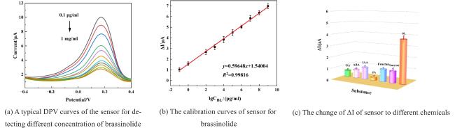
Fig. 6 Performance testing of immunosensors |
According to the approximate proportions of different substances in plants[34-36], the developed sensors were employed to detect various interferences such as gibberellin (GA, 50 ng/mL), jasmonic acid (JA, 4 ng/mL), abscisic acid (ABA, 250 ng/mL), indole-3-acetic acid (IAA, 275 ng/mL), fructose (90 ng/mL), and sucrose (1.4 µg/mL). The results presented in Fig. 6(c) reveal that ΔI for the sensor in response to brassinolide is significantly greater than that observed for of any of the other interfering substances, thus highlighting the sensor's exceptional selectivity for brassinolide.
The same brassinolide solution was detected six times subsequently using the identical electrode, yielding an RSD (Relative Standard Deviation) of ΔI of 2.5% (Fig. 7(a)). To assess the reproducibility across different electrodes, the same concentration of brassinolide solution was analyzed using five separate electrodes, resulting in an RSD of ΔI of 1.9% (Fig. 7(b)). To evaluate the stability of the sensor, it was placed at 4 °C and used to measure the same concentration of brassinolide solution on 2, 4, 7, 10 and 14 days, respectively (Fig. 7(c)). Notably, the ΔI value retained 92.33% of its initial signal after 14 days, suggesting that the immunosensor exhibits satisfactory reproducibility and stability over time.
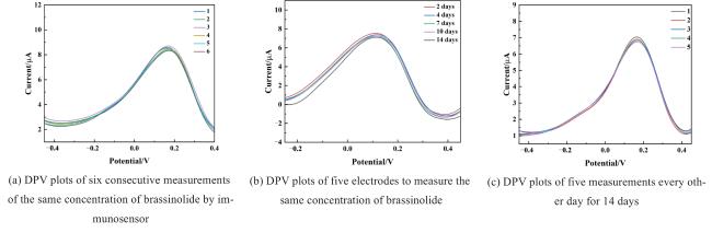
Fig. 7 Stability testing of immunosensors |
2.5 Detection of brassinolide in actual sample
To assess its utility, the developed immunosensor was utilized for the determination of brassinolide in wheat juice. Based on the established calibration curve, the determined concentration of brassinolide in the wheat extract was calculated to be approximately 0.71 ± 0.002 8 pg/mL, aligning well with reported values from previous studies[37]. The recovery rates achieved by the sensor ranges from 98.13% to 104.74% (an shown in Table 2), indicating that the prepared sensor can be applied for the accurate determination of brassinolide in actual samples.
Table 2 Recovery of brassinolide in wheat sample (n = 4) |
| Samples/pg/mL | Added/pg/mL | Found/pg/mL | RSD/% | Recovery/% |
|---|---|---|---|---|
| 0.71±0.002 8 | 1 | 1.68±0.04 | 2.6 | 98.13 |
| 10 | 11.22±0.34 | 3.1 | 104.74 | |
| 100 | 105.42±2.95 | 2.8 | 104.67 | |
| 1 000 | 1 037.67±56.03 | 5.4 | 103.69 |
Due to the flat and thin nature of most plant leaves, traditional electrodes like GCE or needle electrodes are not suitable for direct insertion into the leaves for in situ testing. In this study, a flattened SPE was used as a substrate electrode, providing a suitable fit underneath the plant leaves for in situ detection. The tape was used to fix the electrode below the detection area of leaves. A puncture tool was then used to create small holes in the leaves directly above the working electrode area. Subsequently, 20 μL of PBS buffer was added to the holes, allowing the juice of the leaves could be released onto the working electrode. Following a 30-minute incubation period, a DPV test was performed, and the resulting data can be found in Table 3.
Table 3 The results of BSA/antibody-brassinolide/Mxene@PDA/CuCl2 NWs/AuNPs/SPE for detecting brassinolide in the leaves of wheat seedlings under salt stress (n = 10) |
| Sample | Control | 100 mM NaCl |
|---|---|---|
| Sensor /pg/mL | Sensor/pg/mL | |
| 1 | 9.16 | 17.85 |
| 2 | 9.29 | 13.75 |
| 3 | 8.07 | 19.35 |
| 4 | 7.15 | 18.91 |
| 5 | 6.34 | 19.33 |
| 6 | 8.59 | 16.15 |
| 7 | 6.79 | 14.43 |
| 8 | 6.06 | 18.91 |
| 9 | 9.59 | 17.45 |
| 10 | 10.66 | 20.85 |
| Average | 8.17±1.54 | 17.70±2.29 |
| Significance level | 0.000** | 0.000** |
|
In this experiment, the brassinolide content in the control group is approximately 8.17 ± 1.54 pg/mL, and the content of brassinolide in the salt-stressed group increases to 17.70 ± 2.29 pg/mL, which is about twice that of the control group. There are experiments show that under the plants under salt stress accumulation of some organic molecules such as brassinolide, so as to promote the generation of antioxidant enzymes in plants and reduce the malondialdehyde (MDA) content, to reduce the salt stress the negative effects of plant growth[38]. For example, Lu et al.[39]treated Suaeda glauca with 100 mM NaCl solution, and the concentration of brassinolide was about 1.5 times higher than the control. When Wang et al. [40] treated broccoli sprouts with 80 mM NaCl solutions, the concentration of brassinolide was approximately 2-fold higher than the control. The increase of brassinolide in wheat seedlings under salt stress here can be understood as an adaptive regulation of plants to salt stress[41]. The multiples of the change of brassinolide concentration in plants are consistent with this experimental results, which indicates that the detection results of the immune sensor are accurate and reliable.
3 Conclusions
This work presents an electrochemical sensor based on Mxene@PDA/CuCl2 NWs/AuNPs, which demonstrates high performance in brassinolide determination.
AuNPs are employed to enhance the conductivity and effective active area of the electrode. CuCl2 NWs are also utilized to modify the electrode, not only enhancing its conductivity, but also serving as an electrical signal probe for the immunosensor. Additionally, Mxene@PDA nanocomposite is further coated on the electrode to enhance its conductivity, biocompatibility and antibody adsorption ability. Compared to the previous studies, the developed brassinolide immunosensor exhibits the widest detection range of 0.1 pg/mL~1 mg/mL, and the lowest LOD of 0.015 pg/mL (S/N=3). It was also utilized to detect brassinolide in salt-treated wheat samples in situ, confirming its great potential for in situ studies in precision agriculture.
In summary, this study proposes an electrochemical sensor preparation method that overcomes the limitation of traditional detection and enables in situ detection of small molecules in plants with minimal damage to them.


