


0 引 言
随着物质水平的提高,人们对生活质量有了更高要求,食品安全问题也日益受到重视。当前,国内外市场中许多水果、蔬菜及粮食中都发现有农药残留,这已经成为危害人们健康的一个潜在因素。特别在一些发展中国家,农药中毒成为一个公共卫生问题,每年甚至造成数万人死亡[1]。因此,急需开发快速、灵敏的农药残留检测手段。
目前,农药残留检测方法主要包括气相色谱法[2]、超高效液相色谱法[3]、超高效液相串联质谱法[4]等,但通常受生物样品制备复杂和检测平台的限制。表面增强拉曼光谱散射/光谱(Surface-enhanced Raman Scattering/Spectroscopy, SERS)技术是一种基于拉曼散射现象的分析技术[5]。它可以通过电磁增强机制对吸附特定金属表面上的目标分子的拉曼散射信号进行大幅度增强[6]。SERS检测技术具有检测灵敏度高、指纹特征独特、分析快速等优点[7]。目前,SERS技术已经在食品[8]、医药[9]、农业[10]等各个领域被广泛应用。将SERS技术应用于农药残留检测,可以满足当前农药残留快速检测的需求。
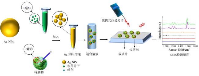
近年来,SERS技术发展很快,其关键在于制备具有SERS活性的纳米材料,其中金/银纳米颗粒(AuNPs/AgNPs)由于在可见光区具有表面等离子体共振吸收,是目前应用较多的SERS材料[11]。一般来说,银纳米粒子的合成方法有柠檬酸钠还原法、盐酸羟胺法等[12]。盐酸羟胺法还原银纳米粒子(AgNPs)的优点是具有较好的形貌可控性、反应速度较快等,但缺点是羟胺是一种致癌物质,对环境污染较大[13];柠檬酸钠还原法的优点是使用环保的还原剂和表面活性剂,操作相对较为安全,制备出的AgNPs形貌规则、分散性好,但缺点是制备过程中需要高温或较长反应时间[14]。为克服上述缺点,本课题组研发了一种常温条件下利用柠檬汁还原的AgNPs的简易方法。该AgNPs不仅具有很好的SERS活性,而且在常温下化学性质稳定,可以保存较长时间。百草枯(Paraquat)化学名称是1-1-二甲基-4-4-联吡啶阳离子盐,是一种快速灭生性除草剂,具有触杀作用和一定内吸作用[15]。虽然百草枯已被绝大多数国家禁用,但仍存在检测需求。多菌灵(Carbendazim)化学名称是2-苯并咪唑氨基甲酸甲酯,是一种内吸性广谱杀菌剂,广泛应用于农业,其产生的残留物持续存在于环境中,对人类和动物健康造成危害[16]。进一步,本研究以检测一些典型农药(如:百草枯和多菌灵)在果蔬上残留为例,验证用此方法制备的AgNPs可以对多种果蔬(如:草莓、西瓜、葡萄、番茄等)其表面农药残留进行微量和快速检测。SERS检测过程见图1。
1 实验方法
1.1 实验试剂与仪器
本实验用的新鲜柠檬购自长丰县水果超市。氢氧化钠(NaOH)、硝酸银(AgNO3)购自中国国药控股化学试剂有限公司。百草枯和多菌灵标准品购自默克(Merck)公司,以上试剂仅用于科研实验。分光光度计,岛津UV-2500,SERS光谱测量范围为200~800 nm。高效液相色谱仪,Agilent 1260。透射电子显微镜(Transmission Electron Microscope, TEM),Talos F200X G2。便携式拉曼光谱仪,如海光电公司研发的RMS1000便携式拉曼光谱仪,配备785 nm激光(功率200 mW),记录光谱范围为400~2 000 cm-1。
1.2 AgNPs的制备
首先将柠檬榨成新鲜柠檬汁,用漏斗过滤,去除柠檬汁中肉眼可见的粗纤维。然后取1 mL的柠檬汁用超纯水稀释成2%的柠檬汁溶液,并配制成一定浓度的AgNO3溶液,以及50 mmol/L的NaOH溶液。最后在室温条件下,向烧杯中分别滴加10 mL超纯水、2 mL NaOH、2 mL柠檬汁及5 mL的AgNO3溶液,静置反应40 min。当溶液颜色由无色变为澄清的黄色时,即可收集放置室温下保存。
1.3 柠檬汁中还原化合物含量的分析
柠檬汁中有多种与AgNPs制备相关的化合物,起主要还原作用的是其中的还原糖及抗坏血酸[17]。柠檬汁中主要还原糖包括葡萄糖和果糖[18],因此,制备浓度分别为20、40、60、80、100 mg/mL的果糖标准溶液和浓度分别为5、10、20、60、100 mg/mL的葡萄糖标准溶液。样品经离心过滤处理后稀释5倍,然后进行紫外光谱的采集。柠檬汁中抗坏血酸(Ascorbic Acid, AA)含量根据文献[19]的方法测定。使用Agilent高效液相色谱进行定量分析。流动相为0.1%的二水合草酸。用流动相分别制备浓度为9.88、19.76、29.64、39.52、49.4 μg/mL的抗坏血酸标准溶液。样品经0.45 μm滤膜过滤后进行色谱定量分析。
1.4 SERS信号的测量
用超纯水配制百草枯标准溶液;用乙醇配制多菌灵标准溶液。根据《食品安全国家标准 食品中农药最大残留限量GB 2763—2021》规定,不同水果中的最低百草枯残留标准0.01 mg/kg,不同水果中最低多菌灵残留标准0.5 mg/kg。因此,制备不同浓度(10-3~10-14 mol/L)的百草枯溶液及不同浓度(10-3~10-10 mol/L)的多菌灵溶液。对于百草枯、多菌灵标准品的SERS检测,分别将不同浓度的百草枯(多菌灵)溶液与AgNPs溶液、NaCl(5%)溶液以体积比1∶2∶1的比例混合,静置1~5 min,在激光波长为785 nm的便携式拉曼光谱仪上进行SERS信号采集。对于实际样品中的农残SERS检测,首先用超纯水反复清洗新鲜水果,然后滴加微量(50—100 μL)不同浓度的待测样品。待农药溶剂完全挥发后,取棉签擦拭果皮表面,将棉签放入装有少量溶剂的离心管中浸泡并超声清洗。最后将获取的样品与AgNPs溶液、NaCl(5%)溶液以体积比1∶2∶1的比例混合进行SERS信号采集。光谱采集时间一般为10~30 s。对获得的SERS光谱进行相关预处理,即对光谱进行Savitsky-Golay平滑处理以减少噪声的影响。此外,使用多项式拟合进行基线校正,以消除荧光背景的影响。峰强度采用平均值±标准偏差(Standard Deviation, SD)进行评估。以上所有操作均在NG-LabSpec仪器软件以及Origin 2023软件中执行。
2 结果与讨论
2.1 AgNPs的制备与表征
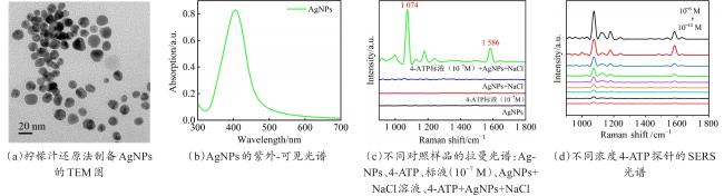
进一步探究AgNPs形貌表征,透射电子显微镜(Transmission Electron Microscope, TEM)结果如图2(a)所示。获得的AgNPs球状结构尺寸均匀,大小一致。随机挑选AgNPs测量,其直径均为20 nm左右。合成的AgNPs的紫外-可见光谱如图2(b)所示,出现一个尖锐的吸收峰,证明AgNPs制备成功。在400 nm附近出现的峰是AgNPs等离子体共振吸收带[20]。为验证所制备的AgNPs的SERS效应,使用4-ATP作为标准探针。首先进行对比测试,如图2(c)所示,分别为AgNPs的拉曼光谱、AgNPs+NaCl的拉曼光谱、4-ATP(10-7 mol/L)的拉曼光谱,以及4-ATP(10-7 mol/L)的SERS光谱。对比发现该纳米粒子及溶液检测体系显示出低且被忽略的背景,并且具有较强的SERS增强效应。由图2(d)可知,由于底物对4-ATP的吸附可实现对其的SERS检测。在1 080 cm-1和1 580 cm-1处4-ATP的特征峰清晰可见,分别对应于C-S和C=C的伸缩振动[21]。该AgNPs可以检测低至10-14 mol/L的探针分子。以上结果均证实AgNPs优异的SERS性能。
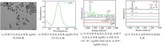
为确定柠檬汁在制备AgNPs中的作用,对柠檬汁主要还原成分进行分析。首先用高效液相色谱测量柠檬汁中抗坏血酸的含量,通过分析抗坏血酸的标准品浓度与峰面积之间的线性关系,得出标准曲线及线性回归方程,由此测出的柠檬汁中抗坏血酸含量。结果显示,所测柠檬汁中抗坏血酸的含量为395.76 μg/mL。此外,利用紫外光谱法测得柠檬汁中果糖、葡萄糖的含量分别为5.95和5.90 mg/mL。进一步,根据各组分的浓度数据,重新配置抗坏血酸、葡萄糖和果糖的混合溶液,由此按上述步骤进行AgNPs再次制备,发现所得AgNPs与原柠檬汁制备的AgNPs在大小形貌上类似。如图3(a)所示,新的AgNPs的大小为20 nm左右,这与柠檬汁合成的纳米粒子大小基本一致,形貌相同,且合成的纳米粒子分布较均匀,其紫外吸收谱同样在400 nm左右出现,证明AgNPs的成功制备(图3(b))。为检验新配的AgNPs的SERS增强效果,对4-ATP重新进行定量检测,图3(c)为AgNPs本身的拉曼光谱及背景光谱,显示其信号峰强低至可以被忽略;图3(d)为4-ATP的SERS定量检测结果,显示4-ATP的检出限为10-8 mol/L,说明柠檬汁还原的AgNPs所测4-ATP灵敏度更高。此外,通过与柠檬酸钠制备的AgNPs进行比较发现,柠檬汁还原的AgNPs的SERS灵敏度更好。
2.2 SERS测量均匀性、稳定性分析
2.2.1 均匀性分析
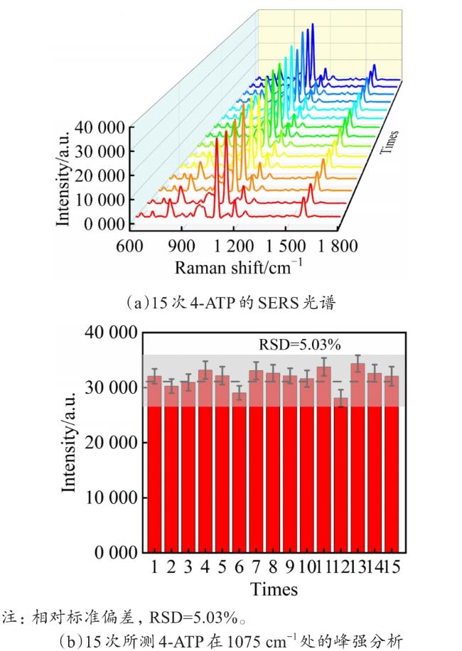
为进一步证明该纳米粒子具有优异的SERS性能,对其均匀性及稳定性进行实验和分析。首先对该纳米粒子的均匀性进行分析。随机采集15次4-ATP(10-7 mol/L)的SERS信号,结果如图4(a)所示,在1 075和1 586 cm-1附近观察到4-ATP的两个较强的特征峰,并且对比15次检测结果,可以发现4-ATP的特征峰强度变化并不明显。
2.2.2 稳定性分析
此外,SERS底物的稳定性对于实际应用也非常重要。为检验该纳米粒子的时间稳定性,将不同储存时长的AgNPs作为底物检测4-ATP(10-7 mol/L)的SERS光谱。所有的SERS底物都被储存在室温(10-7 mol/L)条件下。图5(a)为41 d内不同日期所测的同一浓度4-ATP的SERS光谱比较,可以看到随着时间的增加,4-ATP的拉曼特征峰强度逐渐变弱。由此证明该纳米粒子可以储存时间长约41 d。图5(b)为AgNPs在不同储存时间下检测同浓度4-ATP在拉曼位移为1 076 cm-1处的SERS峰强变化。柱形图与折线图更加直观地展示出随着储存时间的增加AgNPs的SERS效应逐渐降低。但在储存41 d后检测浓度为10-7 mol/L的4-ATPSERS特征峰强度仍较高。上述研究证明该AgNPs有着较好的时间稳定性。
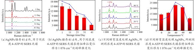
进一步,本研究还检验该纳米粒子的温度稳定性。首先取同体积该纳米粒子10 mL分别在0、20、40、60、80 ℃下孵育30 min;然后将不同温度处理后的AgNPs在室温下进行同一浓度4-ATP的SERS检测;最后分析AgNPs所检测的4-ATP(10-7 mol/L)的拉曼光谱。如图5(c)所示,随着孵育温度的增加,4-ATP的拉曼信号强度呈现先增加后降低的趋势,但总体来说,温度对AgNPs的SERS增强效果影响不明显。因此,可以判定该纳米粒子具有较高的温度稳定性。图5(d)采用柱形图与折线图结合的作图分析方法,分析4-ATP在1 076 cm-1的SERS强度变化,更加直观地显示温度对该纳米粒子SERS增强效果的影响。因此,上述结果表明,该AgNPs具有良好的均一性,长时间常温储存的稳定性及较大范围内的温度稳定性。
2.3 条件优化(pH、AgNO3浓度)
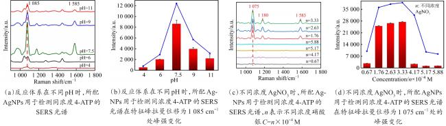
为对加入的AgNO3浓度进行优化,首先保持溶液反应体系的pH为7.5,分别添加浓度为0.67×10⁻⁴、1.76×10⁻⁴、2.63×10⁻⁴、3.33×10⁻⁴、4.17×10⁻⁴、5.17×10⁻⁴、5.88×10⁻⁴ mol/L的AgNO3。将所配的AgNPs用于同浓度4-ATP的SERS检测,如图6(c)所示,可以看到AgNO3浓度为1.76×10-4~3.33×10-4 mol/L时,所配AgNPs的SERS增强效果较好。之后对4-ATP的拉曼光谱在拉曼位移为1 075 cm-1处的SERS峰强进行分析,如图6(d)所示,更为直观地反映出AgNO3浓度为1.76×10⁻⁴~3.33×10⁻⁴ mol/L时所配得的AgNPs具有较强的SERS活性。
3 农残检测实验
3.1 百草枯标准品的SERS检测
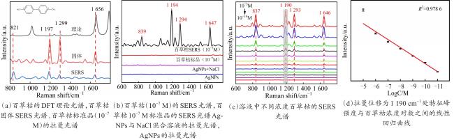
表1 百草枯特征峰分子振动归属表[23]Table 1 The molecular vibration attribution table of paraquat characteristic peaks |
| 拉曼位移/cm-1 | 峰归属 |
|---|---|
| 839 | C-N拉伸 |
| 1 198 | C=C弯曲振动 |
| 1 298 | C-C结构变形 |
| 1 653 | C=N拉伸 |
图7(b)为检测百草枯时的背景信号分析。AgNPs及AgNPs加辅助剂(NaCl)的拉曼峰几乎都为一条直线,说明SERS采集的背景信号可以忽略不计。10-7 mol/L百草枯标准溶液拉曼谱图没有出现特征峰,而10-7 mol/L百草枯的SERS谱图出现较强的特征峰信号,说明该AgNPs具有很好的SERS活性。如图7(c)所示,用该纳米粒子所测百草枯的检测限可以低至10-14 mol/L。对所测百草枯进行定量分析(图7(d)),浓度为10-5~10-11 mol/L的百草枯SERS光谱中,在拉曼位移为1 190 cm-1处的特征峰强度与百草枯浓度的对数呈现良好的线性关系。说明该SERS基底可以用于百草枯的定量检测。
3.2 多菌灵标准品的SERS检测
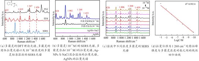
表2 多菌灵特征峰分子振动归属表[24]Table 2 Molecular vibrational attribution table of carbendazim characteristic peaks |
| 拉曼位移/cm-1 | 峰归属 |
|---|---|
| 620 | C-C-C面内弯曲振动 |
| 1 017 | C-N弯曲和C-弯曲及C-O-CH3拉伸 |
| 1 266 | C-H弯曲和N-H弯曲 |
| 1 454 | C-H弯曲和N-H弯曲 |
图8(b)为检测多菌灵时的背景信号分析。AgNPs及AgNPs加辅助剂(NaCl)的拉曼峰几乎都为一条直线,说明SERS采集的背景信号可以忽略不计。10-7 mol/L多菌灵标准溶液拉曼谱图没有出现特征峰,而10-7 mol/L多菌灵的SERS谱图出现较强的特征峰信号,说明该AgNPs具有很好的SERS活性。如图8(c)所示,用该纳米粒子所测多菌灵的检测限可以低至10-10 mol/L。对所测多菌灵进行定量分析(图8(d)),浓度为10-4~10-10 mol/L多菌灵的SERS光谱中,在拉曼位移为1 268 cm-1处的特征峰强度与多菌灵浓度的对数呈现良好的线性关系。说明该SERS基底也可以用于多菌灵的定量检测。
3.3 果蔬表皮上百草枯、多菌灵农残的SERS检测
进一步,本研究对果蔬上的农残进行SERS测量验证。在实验中,选取草莓、番茄、西瓜和葡萄等果蔬,将配制好的一系列浓度的百草枯、多菌灵标准溶液滴加到果皮上,待溶液完全挥发之后,用棉签蘸取表皮农药残留。将收集到的农残放入溶剂中,在超声中振荡清洗,之后取上清溶液进行农残SERS信号的采集。图9(a)~图9(d)分别为百草枯处理西瓜、番茄、葡萄和草莓表皮后采集的农残SERS光谱。经农药处理后的西瓜、番茄、葡萄、草莓四种果蔬表皮均可以检测到在836、1 190、1 294、1 647 cm-1处的百草枯农药特征峰。这与标准品SERS光谱结果一致。图9分别为西瓜、番茄、葡萄和草莓上百草枯残留的表面增强拉曼光谱。
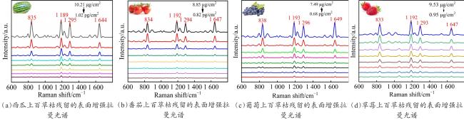

计算结果显示,果蔬实际样品中的百草枯最低检测浓度分别为1.02(西瓜)、0.85(番茄)、0.68(葡萄)和0.95 pg/cm2(草莓),换算成单位质量果蔬的农残含量,最低检测限可达3.90 ng/kg。多菌灵的最低检测浓度分别为47.82(草莓)和44.60 pg/cm2(苹果),换算成单位质量果蔬的农残含量,最低检测限可达0.22 μg/kg。表3为SERS检测百草枯、多菌灵的检出限与农药残留检测限(Limit of Detection, LOD)国标对比,可见这些检测值都远低于《食品安全国家标准 食品中农药最大残留限量(GB2763—2021)》规定的最高残留水平。以往报道百草枯的检测限可以达到0.5 ng/kg,检出时间为15 s[23],多菌灵的检出限可以达到0.85 µg/kg,检出时间为10 s[25]。本研究两种农残检出时间均为10 s。
4 结 论
综上所述,本研究报道了一种基于柠檬汁还原法制备AgNPs的简易方法。该方法具有绿色环保、方便快捷的优点,所制备的AgNPs形貌均匀,具有很好的SERS活性,并且化学性质稳定,可以在常温下长期储存。此外,本研究还明确了柠檬汁中主要还原成分抗坏血酸、葡萄糖、果糖的含量及在制备银纳米颗粒中的作用。利用该方法制备的AgNPs,对草莓、西瓜、葡萄和番茄等果蔬表皮上百草枯、多菌灵等农残进行了SERS检测,可得其浓度检测限达到1~50 pg/cm2,农残检测限LOD远低于最大残留量(Maximum Residual Level, MRL)。这项工作为农产品的农残微量检测提供了快速、无损、便捷的方法。


